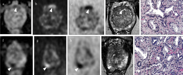
Figure 4. a–l.
Biparametric MRI of the prostate at 3T in a 71-year-old patient with PSA=11 ng/mL with multifocal prostate cancer. Ovalar lesion located in the anterior transition zone at midgland is assigned to S-PI-RADS category 3b lesion (volume>0.5 cc, targeted biopsy is indicated); (arrowhead in a) the lesion is moderately hypointense on axial ADC map, (arrowhead in b) hyperintense on axial DWI at high b-value, (arrowhead in c) hypointense on axial DWI at high b-value inverted, and (arrowhead in d) hypointense on axial T2WI: (e) Gleason score 6 on histology after targeted TRUS/MRI transperineal biopsy. The smallest round (index lesion) in the peripheral posteromedial zone at the apex is assigned to S-PI-RADS category 4 lesion (volume<0.5 cc, targeted biopsy is indicated); (arrowhead in f) the lesion is markedly hypointense on axial ADC map, (arrowhead in g) hyperintense on axial DWI at high b value, (arrowhead in h) hypointense on axial DWI at high b-value inverted, and (arrowhead in i) hypointense on axial T2WI: (l) Gleason score 7 on histology after targeted TRUS/MRI transperineal biopsy.

