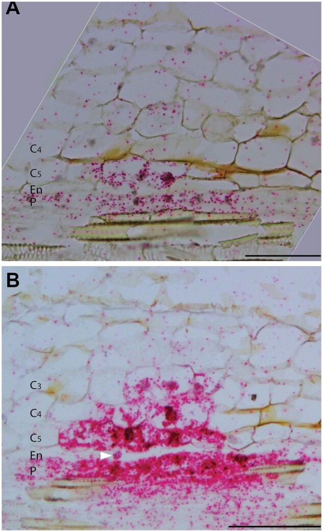
Figure 5.

MtHDT2 is expressed in nodule primordia. In situ hybridization pattern of MtHDT2 mRNA in nodule primordia at stage I (A) and stage III (B). Longitudinal sections of wild-type nodule primordia are shown. Red dots are hybridization signals. Divided and dividing primordium cells are distinguished by their smaller size (10–60 µm) than nonprimordium cells (>100 µm). Arrowhead in (B) indicates a nucleus from an endodermal cell that has not divided. P, Pericycle; En, Endodermis; C5/4/3, the fifth/fourth/third cortical cell layer. Scale bars = 100µm.
