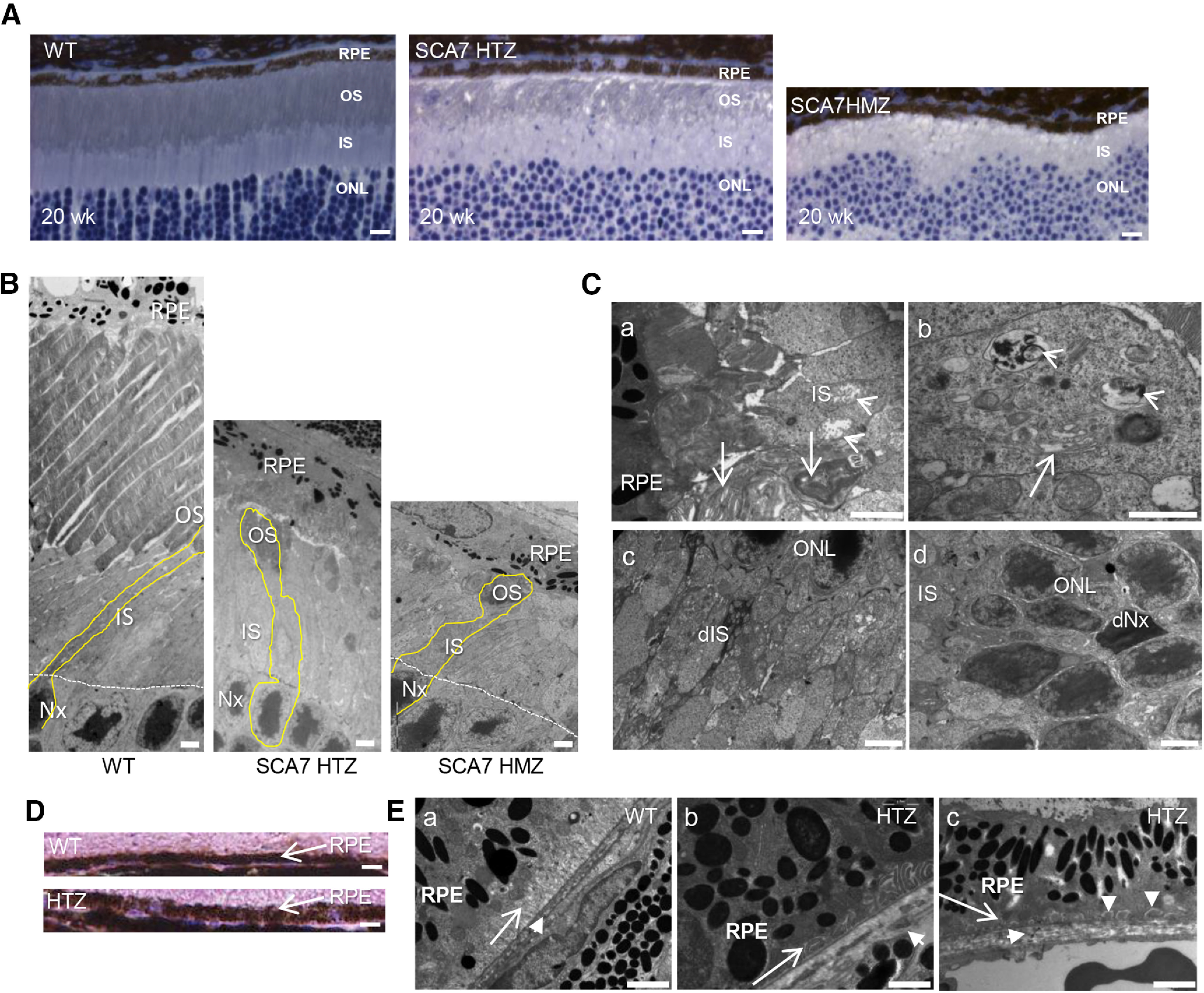
Figure 3.

Tissular and cellular alterations of SCA7 mouse eye. A, Histologic sections of retina at 20 weeks represent the progressive thinning of outer segment (OS) layer in the SCA7140Q/5Q heterozygous (HTZ) retina and the absence of OS in the more severe SCA7140Q/140Q homozygous (HMZ) retina compared with WT retina. IS, Inner segment layer. Scale bar, 15 µm. B, Electron micrographs represent the progressive disappearance of OS and the shortening of IS of 44-week-old SCA7 HTZ and 20-week-old SCA7 HMZ, compared with WT mice. Photoreceptor nuclei (Nx). Scale bar, 10 µm. C, Electron micrographs of 20-week-old SCA7 HMZ retina. The remnant OSs have lost their parallel organization (arrows), and the IS contains swollen and disrupted mitochondria (arrowheads) (Ca). The IS shows dilated and vesiculated endoplasmic reticulum (arrow) and accumulation of vesicular membranes (arrowhead) (Cb). Dark degenerating IS (dIS) with numerous abnormal mitochondria (Cc). Dark photoreceptor nuclei (dNx) (Cd). Scale bars: Ca, Cb, 2 μm; Cc, Cd, 5 µm. D, Histologic sections comparing the thickness of WT and SCA7 RPE at 20 weeks. Scale bar, 15 µm. E, Electron micrographs comparing the basal infolding membrane of RPE and Bruch's membrane of WT and SCA7 mice. In WT (Ea), the basal infolding membrane of RPE (arrow) shows a typical lamellar organization with translucid lumen, and a regular Bruch's membrane (short arrow). In contrast, in SCA7 retina, the infolding membrane (long arrow) is either opaque (Eb) or completely absent (Ec), whereas the Bruch's membrane (short arrow) is enlarged and disorganized (Eb, Ec), and occasionally interrupted (Eb). In addition, homogeneous deposits are found between the basement membrane and plasma membrane of RPE (vertical arrowheads in Ec). Scale bar, 2 µm.
