Figure 6.
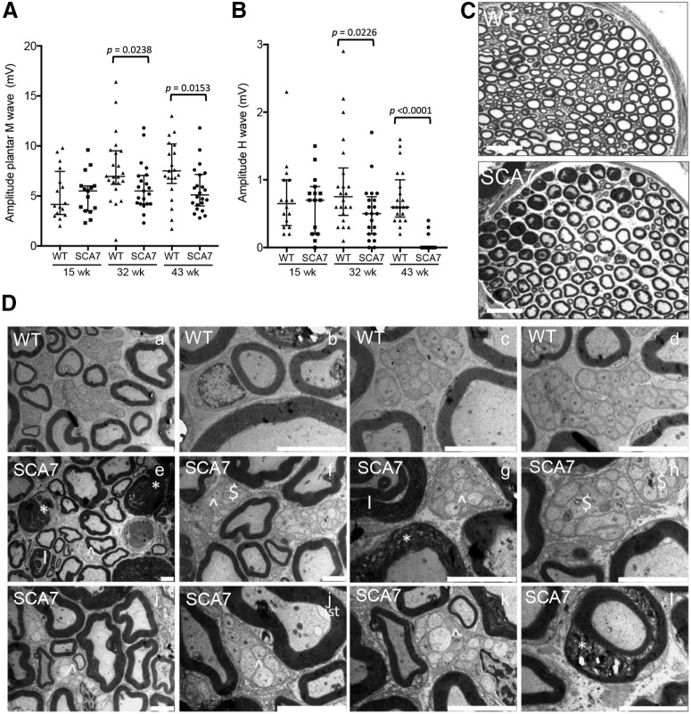
Peripheral nerve alterations in SCA7 mice. Electromyograph activities of the sciatic nerve. Amplitude of motor M-wave (A) and sensory H-wave (B) in SCA7140Q/5Q mice relative to WT littermates (15 weeks: n = 16 WT and 15 SCA7; 32 week: n = 22 WT and 21 SCA7; 43 week: n = 22 WT and 21 SCA7). Scatter plot represents median with interquartile range. Mann–Whitney test. C, Comparison of semithin sections of sciatic nerve of SCA7140Q/5Q and WT mice. The mutant shows irregular and degenerated myelinated fibers and loss of small fibers. Scale bars, 50 µm. D, Electron microscopy analyses of the sciatic nerve. Compared with WT littermates (Da-Dd), sciatic nerve section of SCA7140Q/5Q mice (De-Dl) at 43 week shows severe abnormalities in both myelinated and nonmyelinated fibers, including myelin degeneration (* in De, Dg, Dl), infolding-like structure (I in De, Dg), abnormal Remak bundle (∧ in De-Dg, Di-Dk), autophagy ($ in Df, Dh), and inner swelling tongue (ist in Dj). Scale bars: Da, Dj, Dk, 5 µm; Db-Dd, Df-Di, Dl, 2 µm; De, 10 µm.

