Figure 5.

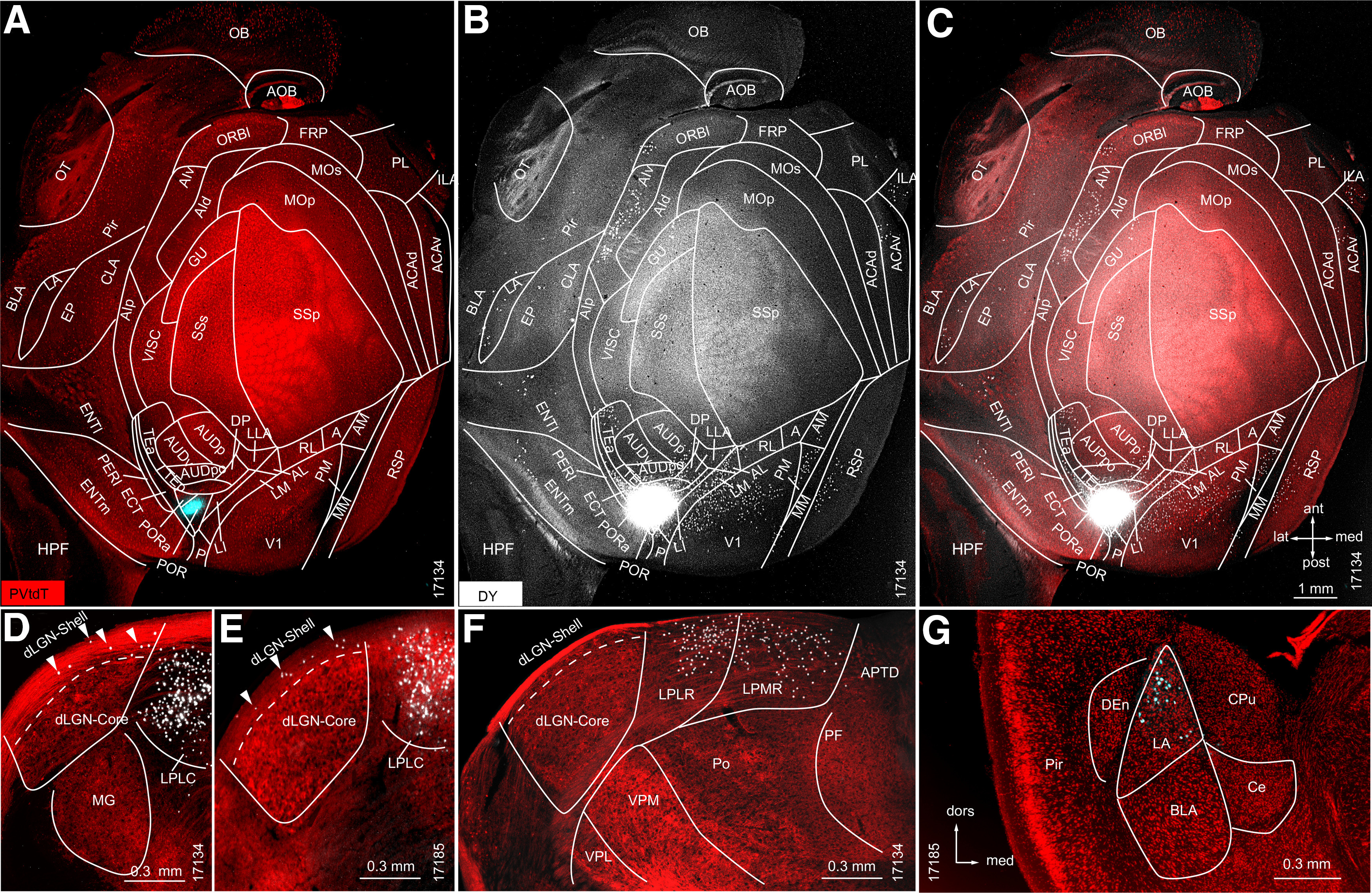
Retrograde tracing in PVtdT mice of subcortical and cortical source neurons projecting to POR. A, Tangential section through L2/3/4 of flatmounted PVtdT-expressing (red) cortex, shows DY injection site (false colored blue) in POR. B, Overexposed black/white image (notice the artificially large injection site gives the false impression that DY spilled beyond the borders of POR) showing DY-labeled neurons (white dots) in LA and multiple cortical areas segmented according to Gămănuţ et al. (2018). C, Overlay of DY-labeled neurons with PVtdT expression. D–G, Coronal sections through thalamus and amygdala. D, E, Representative sections through the dLGN of two different mice (17134 and 17185) showing that DY-labeled neurons are present in the shell (arrowheads) but not in the core. F, G, Dense labeling in the LPLR and LPMR (F), and LA (G).
