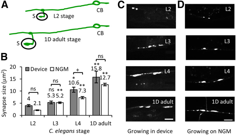
Figure 7.
High-resolution imaging of GFP::RAB-3 in a developing C. elegans inside the microfluidic device. A, Schematic of PLM neuron with its cell body (CB), its neuronal process, and its ventral synapse (S) at L2 larva and 1D adult stages. B, The plot of synapse sizes at different developmental stages of C. elegans growing inside a microfluidic device and on NGM plates. Data represented as mean ± SEM. Statistical significance was evaluated by one-way ANOVA with Bonferroni post hoc comparisons; *p < 0.05 and **p < 0.005 when compared with data from L2 stage; +p < 0.05 and ++p < 0.005, ns, p < 0.05 when compared between animals grown in the device and on NGM solid media; nonsignificant comparisons are not indicated. The number of synapses analyzed from animals grown on NGM solid media is n ≥ 37 and in devices are n = 18 (L2), 16 (L3), 14 (L4), and 8 (1D adult). C, D, Images of PLM synapses at L2, L3, L4, and 1D adult stages of development grown in the microfluidic device (C) and on NGM solid media plates and imaged on agar pads (D). Scale bar: 10 μm.

