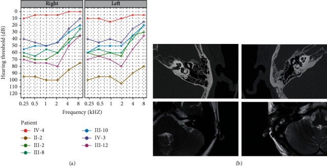
Figure 2.

Audiological and imaging evaluation. (a) The PTA of the right and left ears of representative affected family members and a normal family member. (b) Internal auditory canal MRI and CT scans of the temporal bone in the proband (III-2). The first line contains two CT images of the proband's bilateral temporal bones. The second row contains two MR images of the proband's bilateral internal auditory canals.
