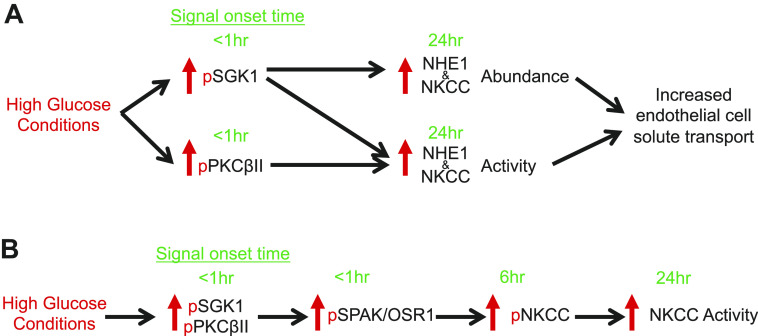
Figure 7.
Hypothesized signaling pathway for high glucose effects on blood-brain barrier endothelial cells. Schematic illustrating the time courses of high glucose effects on blood brain barrier endothelial SGK1, PKCβII, SPAK/OSR1, NKCC and NHE1. Green text highlights the time of onset for observed changes in phosphorylation of the kinases as well as increases in NKCC and NHE1 abundance activity. Arrows indicate downstream activation with likely involvement of intermediate signaling molecules/kinases. A: high glucose exposure causes a rapid transient phosphorylation (a measure of activation) of SGK1 and PKCβII that occurs within 5 min followed by increases in NKCC and NHE1 abundance and activity observed by 24 h. B: high glucose also causes a rapid phosphorylation SPAK/OSR1 (a measure of activation) that is observed by 5 min and persists through 24 h with a subsequent phosphorylation of NKCC at 6 hours that is blocked by inhibition of SPAK/OSR1. Note that our previous studies observed significant increases in NKCC activity (evaluated as bumetanide-sensitive K flux) after 24 h of high glucose, with a trend of increased activity after 6 h albeit one that did not reach statistical significance. Here, we observed significant increases in NKCC phosphorylation starting after 60 min.

