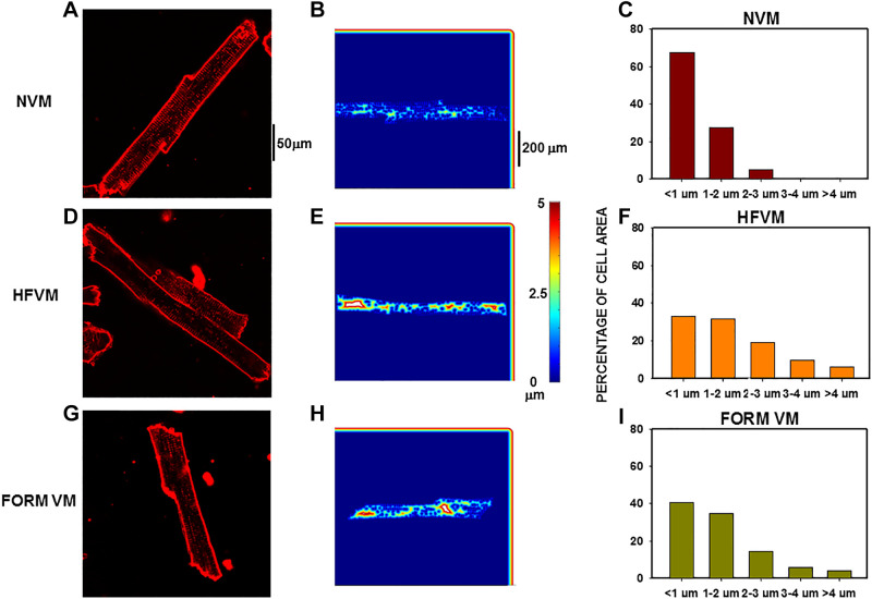
Figure 2.
Distance mapping in normal, heart failure (HF), and formamide (FORM) detubulated dog ventricular myocytes (VMs). Nearest neighbor analysis for distance to closest sarcolemmal or t-tubule membrane from each pixel in the cell interior. A and B: confocal image of a normal VM (NVM) (A) with the distance plot for that cell (B). C: distribution of al pixel distances for this myocyte. D–F: similar presentations for a HFVM. G–I: similar presentations for a FORM myocyte.

