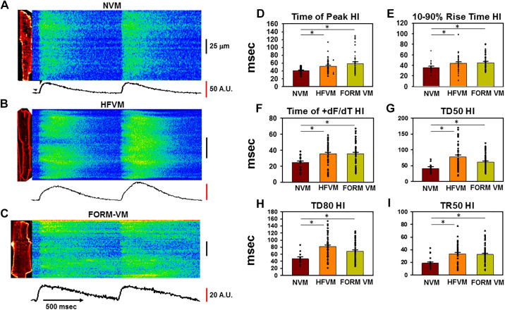
Figure 4.
Defects in intracellular Ca2+ release variability [heterogeneity index (HI)] following TT remodeling in heart failure (HF) and formamide (FORM)-treated dog ventricular myocytes (VMs). A–C: confocal linescan images of Ca2+ transients during pacing at 2 Hz in a normal VM (NVM; A), HFVM (B), and FORM-treated myocyte (C). A.U., arbitrary units. The corresponding 2-dimensional image is shown to the left of each linescan. Average fluorescence along the length of the line is shown below each image. D–I: summary data showing HI for variability in time to peak, rise time, time of maximum dF/dt, duration at 50% of peak (TD50), transient duration at 80% of recovery (TD80), and time to 50% of release (TR50). *P < 0.05, as measured using a single-tailed ANOVA with a Dunn’s test for secondary comparisons; n = 19-20 myocytes in 4 dogs (NVM), 42 in 4 dogs (HFVM), and 49 in 6 dogs (FORM VM).

