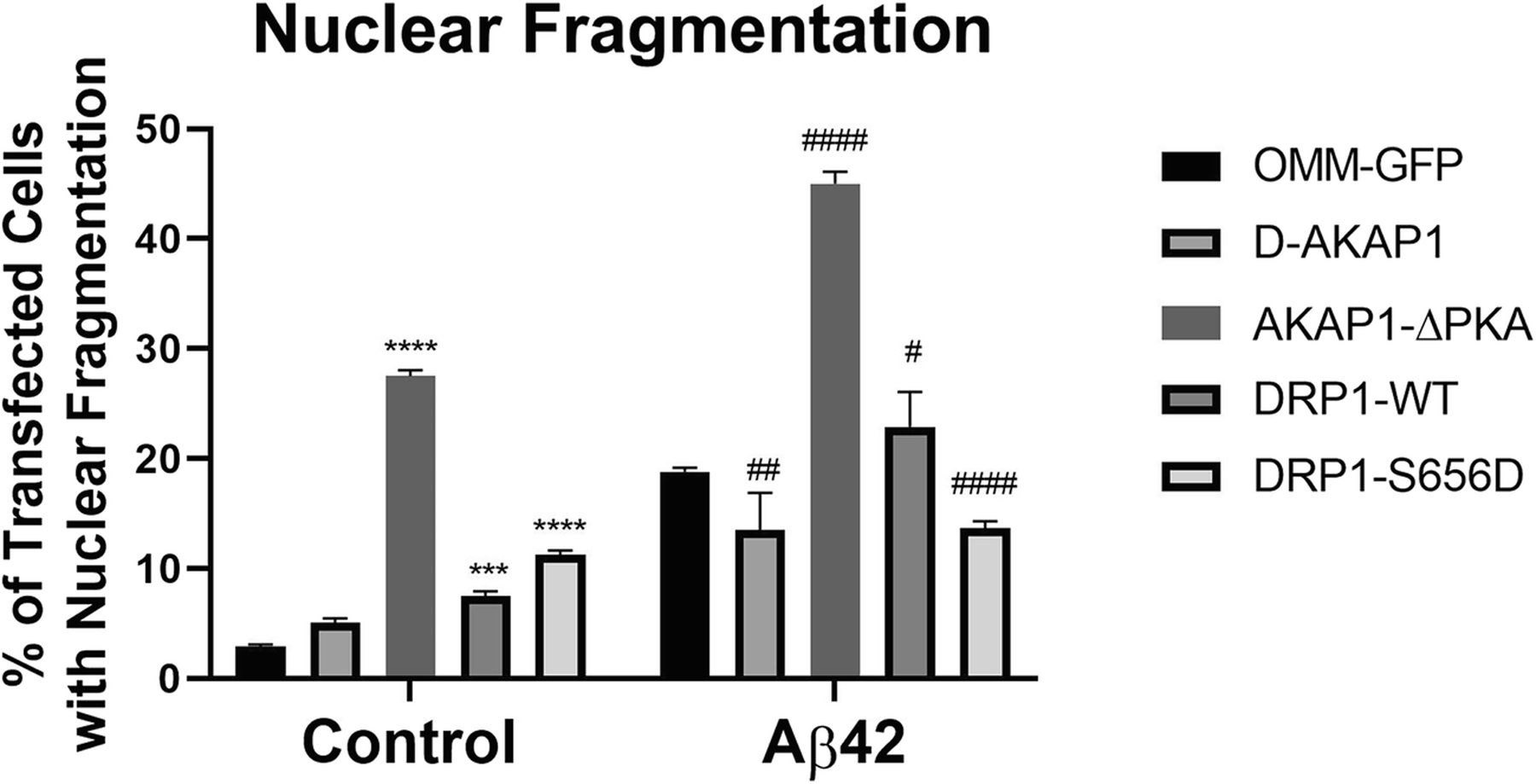
Fig. 4.

D-AKAP1/PKA decreases neuronal apoptosis induced by Aβ42 via PKA-mediated phosphorylation of Drp1.
Bar graph showing a compiled quantification of the percentage of GFP-positive primary cortical neurons containing fragmented or pyknotic nuclei. Primary cortical neurons were treated with vehicle or Aβ42 (24 hr., 10μM). Means ± SEM, derived from an average of 226 primary cortical neurons per construct from at least 10 epifluorescence microscopic fields per experiment, were compiled from three independent experiments (****:p<0.0001 vs. OMM-GFP, ##/###/####:p<0.01 vs. OMM-GFP/Aβ42; Two-Way ANOVA with Tukey’s post hoc test).
