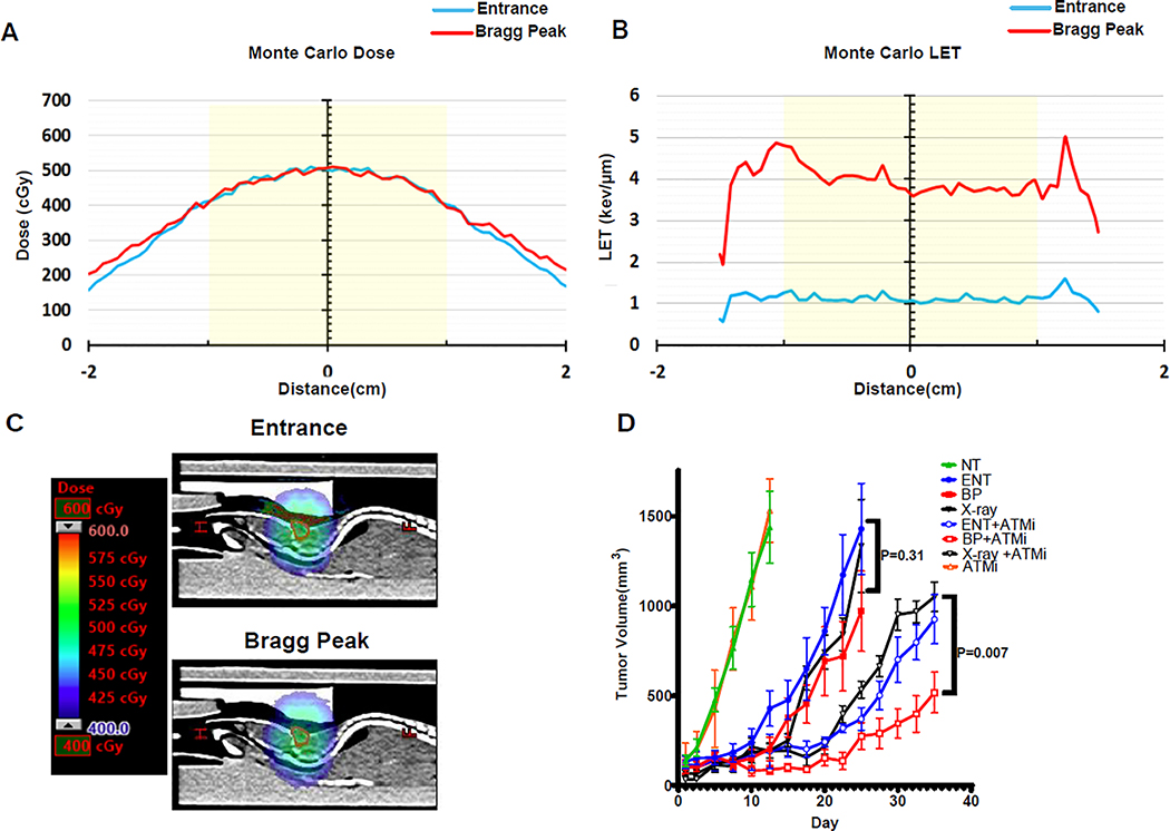
Figure 5. ATM inhibition promotes the efficacy of Proton Bragg peak irradiation in vivo.
A-D MC-BR-BTY-0030 triple negative breast cancer patient-derived xenograft cells were subcutaneously injected into the midline of athymic nude mice. After tumors reached 50mm3 animals were randomized to sham (no treatment [NT]), or 5 Gy (500 cGy) delivered with 6 MV photons (X-ray), 76.8 MeV protons at the entrance (ENT) portion of the Bragg curve, or 76.8 MeV protons at the middle of a 1.5 cm wide spread-out Bragg peak (BP or LET-optimized protons) in a single fraction, with or without pre-treatment one hour prior with the ATM inhibitor, AZD0156 (10mg/kg). AZD0156 was also administered a second time at 24 hours. A, Monte Carlo dose calculation for the entrance and Bragg peak proton plans showing the same physical dose administered. B, Monte Carlo LETd calculation for the entrance and Bragg peak proton plans. An approximate 4-fold increase in LETd with the Bragg peak plan was achieved. Yellow shade in A and B denotes a 2 cm diameter easily encompassing the tumor. C, Sagittal CT image from the entrance and Bragg peak proton plans demonstrating the same physical dose administered, as in A. The tumor volume is delineated in red. D, Tumor volume was assessed over time with day 1 denoting the first dose of AZD0156 and day of irradiation. Shown are the representative data (mean ± SEM) from biologically independent samples (n=7). P values are displayed for the comparison of photons and Bragg peak protons obtained using the two-sided unpaired t-test.

