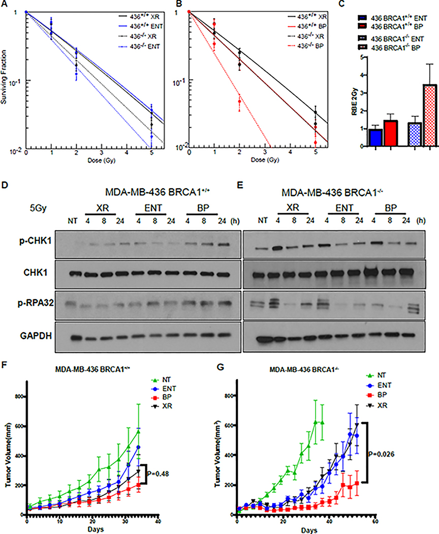
Figure 6. The RBE of proton Bragg peak irradiation is enhanced in BRCA1 deficient tumor cells.
A-B, MDA-MB-436 BRCA1+/+ and BRCA−/− sub-lines were irradiated with 6MV photons or 76.8 MeV protons delivered at the entrance (ENT) or the Bragg peak (BP) of the proton beam profile and clonogenic survival was assessed, (mean ± SEM) from three independent experiments are plotted. C The proton entrance and Bragg peak RBE2Gy for the experiments in A-B was quantified. D, E MDA-MB-436 BRCA1+/+ and BRCA1−/− cells were irradiated with 5 Gy using 6 MV photons(X-ray, XR) or 76.8 MeV protons delivered at ENT or the BP. Lysates were collected at the indicated time points and Western blot analysis was performed using the indicated antibodies. Tubulin was used as a loading control. F,G MDA-MB-436 BRCA1+/+ cells (D) or MDA-MB-436 BRCA1−/− cells were subcutaneously injected into the midline of NOD-SCID mice. After tumors reached 50mm3 animals were randomized to sham (no treatment [NT]), or 5 Gy delivered with 6 MV photons (XR), 76.8 MeV protons at entrance (ENT), or 76.8 MeV protons at the Bragg peak (BP) (LET-optimized protons) in a single fraction. Tumor volume was assessed over time. Shown are the representative data (mean ± SEM) from biologically independent samples (n=9). P values are displayed for the comparison of x-rays and Bragg peak protons obtained using the two-sided unpaired t-test.

