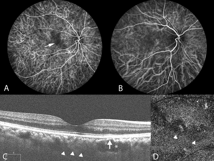
Figure 1.
Multimodal imaging of the right eye of a 45-year-old man with chronic central serous chorioretinopathy. Pachyvessels and attenuation of the inner choroid are seen. (A) Indocyanine green video angiography image in the early phase revealing expanded hypofluorescence (white arrow), indicating a hypoperfused capillary segment. ICGA images revealing that the expanded hypofluorescence was colocalized with underlying pachyvessels (B) and choriocapillaris attenuation (D). (B) ICGA image indicating dilated choroidal vessels near the macula. Pachyvessels are shown as dilated choroidal vessels that do not taper toward the posterior pole. (C) Subretinal fluid resolved at the 4-month follow-up. Enhanced-depth imaging optical coherence tomography (OCT) demonstrated pachyvessels as larger hyporeflective lumen (white asterisk) and obliteration of overlying Sattler’s layer and choriocapillaris (white arrow). The inner surface of the sclera was demarcated, as indicated by the white arrowheads, and the subfoveal choroidal thickness was 315 μm. (D) Attenuation of flow signal within the choriocapillaris (white arrowheads) on OCT angiography.

