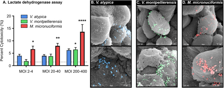
Fig. 1. M. micronuciformis and V. montpellierensis induces significant cytotoxicity in 2-D cervical cell monolayers compared to V. atypica.
A LDH assay to quantify cytotoxicity in cervical epithelial monolayers. M. micronuciformis infections induced significant cytotoxicity to cervical epithelial monolayers at a range of MOIs from 2 to 400. V. montpellierensis infections induced significant cytotoxicity at the highest MOI only. Error bars designate standard deviation. Statistical differences were tested using one-way ANOVA with Dunnett’s adjustment for multiple comparisons, *P < 0.05, **P < 0.005 ****P < 0.0001. Scanning electron microscopy (SEM) images of B V. atypica, C V. montpellierensis, and D M. micronuciformis colonizing 3-D cervical epithelial cells. SEM images were pseudo-colored using Photoshop 19.0 CC (Adobe).

