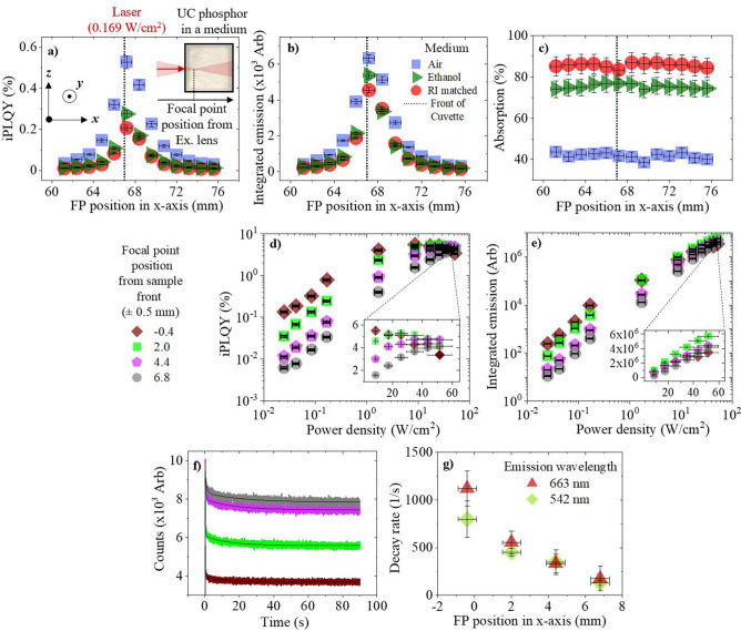
Figure 4.
β-NaYF4:(18%)Yb3+,(2%)Er3+ phosphor is excited with a 980 nm laser diode, the FP of which, is moved through the sample in the x-axis. The phosphor is air (blue square), ethanol (dark green triangle), or a RI matched medium (red circles). The following is measured against the FP position from the excitation lens: (a) iPLQY (500–700 nm), with an inset depiction of the excitation conditions inside each sample, (b) integrated emission (500–700 nm), and (c) absorption. The second experiment investigates moving a high PD regime, where the FP is moved to four positions: − 0.4 (rotated brown square), 2 (light green square), 4.4 (pink pentagon), and 6.8 ± 0.5 mm (grey circle), relative to the front of the cuvette. At various FP positions through the sample, the following is presented against excitation PD: (d) iPLQY (500–700 nm), with an inset magnification of results at the highest PDs, and (e) the integrated emission (500–700 nm), with an inset magnification of the results at the highest PDs. The third experiment investigates excitation induced thermal effects at various FP positions. At various FP positions through the sample, the following is obtained: (f) the 542 nm emission decay versus excitation time, and (g) the 542 nm (rotated green square) and 663 nm (dark red triangle) emission decay rate versus FP position.

