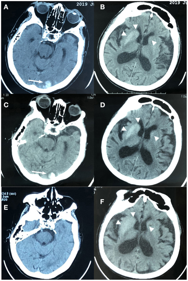
Figure 1.

Brain computed tomography (CT) imaging of an 88-year-old woman with non-ketotic hyperglycemia chorea-ballism and intracerebral hemorrhage. (A,B) At admission, CT showed hyperdense lesions in the left occipital lobe (white arrow), right caudate nucleus and putamen (triangle), and left caudate (triangle). (C,D) Eight hours later after admission, CT showed mild hematoma enlargement, slightly increased edema around the occipital lobe lesion (white arrow), and no significant changes in the bilateral striatum regions (triangle); (E,F) 15 days before admission, CT showed hyperdensity in bilateral striatum regions (triangle) without edema and occipital lobe lesion.
