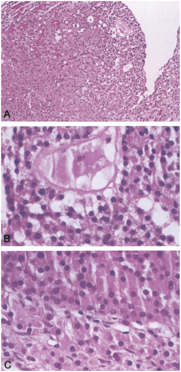
Figure 1.

(A) Luteinized follicle containing a structure suggestive of an ovum undergoing degeneration (hematoxylin and eosin stain; magnification, ×150). (B) Higher magnification of the putative ovum (hematoxylin and eosin stain; magnification, ×250). (C) Higher magnification of the same follicle showing luteinized cells characterized by their larger size, abundant eosinophilic cytoplasm, and prominent nucleus (hematoxylin and eosin stain; magnification, ×250).
