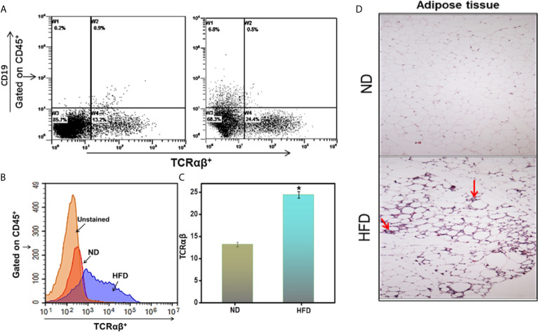
Figure 2.
DIO induces immune cell infiltration in the adipose tissue. Peripheral AT was isolated from the two groups of mice (HFD and ND) after 16 weeks and stained for CD45+, CD19, and TCRαβ+ Abs. (A) Shows a representative experiment indicating the percentages of TCRαβ+ in the cells gated on CD45+ in peripheral fat adipose tissue. (B) Shows a representative overlay histogram of TCRαβ+. (C) Shows cumulative data represent the total percentage of cells ± SEM; total n = 18 (six mice per group) from three independent experiments. The statistical significance between ND and HFD is assessed by Student’s t-test. *p < 0.05 indicate statistically significant differences in TCRαβ+ between ND and those fed HFD. (D) Shows adipose tissue was isolated and fixed in 4% paraformaldehyde, embedded in paraffin, and sectioned at 6 μm. Sections were stained with hematoxylin and eosin. Sections were examined microscopically at a magnification of 10X by a blinded investigator not related to this study. The mice that received HFD had increased the size of adipose cells and marked leukocyte infiltration (indicated by arrow).

