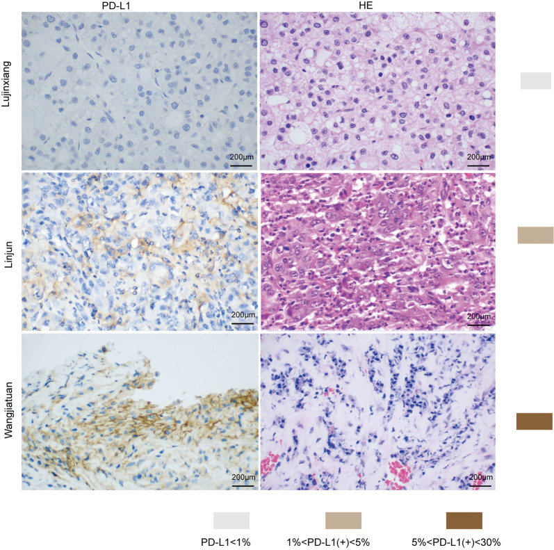
Figure 1.
PD-L1 expression in HCC tissue samples. (A) PD-L1 immunohistochemistry of Liujinxiang HCC biopsy. (B) HE staining of Liujinxiang HCC biopsy. (C) PD-L1 immunohistochemistry of Linjun HCC biopsy. (D) HE staining of Linjun HCC biopsy. (E) PD-L1 immunohistochemistry of Wangjiatuan HCC biopsy. (F) HE staining of a representative HCC biopsy. HCC, hepatocellular carcinoma. Scale bars =200 µm.

