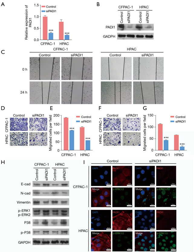
Figure 2.
The knockdown of PADI1 suppressed PAAD cell migration and invasion, and activated the ERK1/2-p38 signaling pathway. CFPAC-1 and HPAC cells were transfected with siPADI1 or a control. (A) A quantitative real-time PCR and (B) western-blot analysis were performed to determine the expression of PADI1 mRNA and protein levels in the above two cell lines. (C) A wound-healing assay was performed to assess cell migration in the above two cell lines (40×). A Transwell assay was used to evaluate cell migration (D,E) and invasion (F,G) in the above two cell lines (100×, stained with crystal violet). Data are expressed as means ± SD from three independent experiments. ***, P<0.001, compared with the control. (H) The protein expression of E-cadherin, N-cadherin, Vimentin, p-ERK1/2, p38, and p-p38 was measured in the above two cell lines by a western-blot analysis. (I) PADI1 and p-ERK1/2 expression levels were observed using immunofluorescence (400×) in the above two cell lines. PADI1, peptidylarginine deiminase 1; PAAD, pancreatic ductal adenocarcinoma; PCR, polymerase chain reaction; mRNA, messenger RNA; SD, standard deviation.

