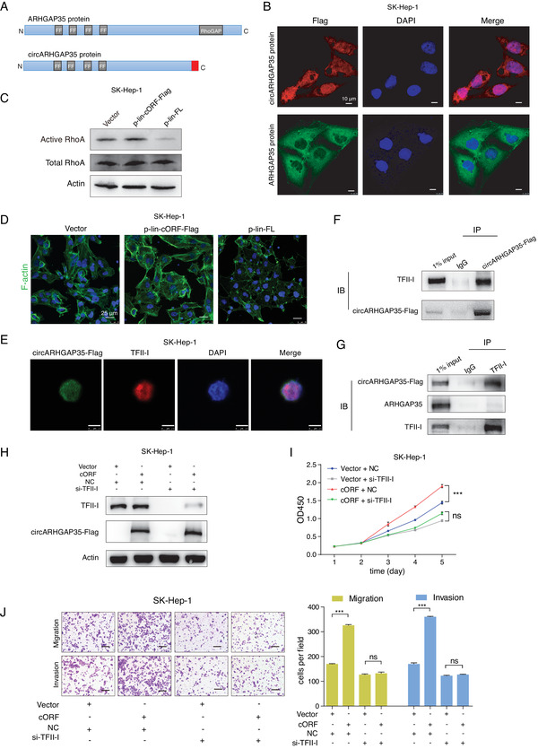
Figure 5.

circARHGAP35 protein interacts with TFII‐I in the nucleus. A) Schematic illustrations of the protein domains of ARHGAP35 and circARHGAP35 proteins. The distinct C‐terminus of circARHGAP35 is shown in red. B) Subcellular localizations of circARHGAP35 protein and ARHGAP35 protein in SK‐Hep‐1 cells infected with circARHGAP35 expressing lentivirus or linear ARHGAP35 expressing lentivirus. circARHGAP35 protein (secondary antibody, Alexa 633, red), ARHGAP35 protein (secondary antibody, Alexa 488, green), and DAPI (blue). Scale bars, 10 µm. C) Western blot analysis of active and total RhoA in SK‐Hep‐1 cells with stable overexpression of circARHGAP35 protein, or ARHGAP35, or vector control. D) F‐actin was stained using phalloidin‐488 in SK‐Hep‐1 cells with stable overexpression of either circARHGAP35 protein or ARHGAP35 protein. Nuclei were stained with DAPI. Scale bars, 25µm. E) Immunofluorescence of circARHGAP35 protein and TFII‐I using Flag or TFII‐I antibody in SK‐Hep‐1 cells infected with circARHGAP35 expressing lentivirus. circARHGAP35 protein (secondary antibody, Alexa 488, green), TFII‐I (secondary antibody, Rhodamine, red), and DAPI (blue). Scale bars, 7.5 µm. F) Immunoprecipitation (IP) assay in SK‐Hep‐1 cells with stable overexpression of circARHGAP35 protein using either Flag or control IgG antibody, followed by immunoblotting using the TFII‐I antibody. G) IP assay in SK‐Hep‐1 cells with stable overexpression of circARHGAP35 protein using either TFII‐I or control IgG antibody, followed by immunoblotting using indicated antibodies. H) Western blot validation of circARHGAP35 protein and TFII‐I protein in circARHGAP35 protein overexpressing SK‐Hep‐1 cells following transfection with siRNA targeting TFII‐I. I,J) CCK‐8 proliferation (I) and transwell (J) assays following transfection with siRNA targeting TFII‐I in circARHGAP35 protein overexpressing SK‐Hep‐1 cells. Scale bars, 10 µm. Data were represented as mean ± SEM. Two‐way ANOVA and Tukey post hoc test were performed for (I); one‐way ANOVA and Dunnett post hoc test were performed (J), ***p < 0.001.
