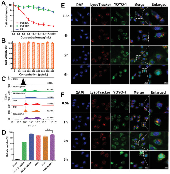
Figure 2.

Evaluation of cytotoxicity, cellular uptake, and endosome escape ability. The cellular viability of B16‐F10 cells after being treated by A) PEI 25K, PEI 1.8K, PR and B) HRMP polymer. C) The cellular uptake analysis of PEI 1.8K/pDNA, PEI 25K/pDNA, core, PUN, and PUN+MMP‐2 using flow cytometry and D) quantitative analysis of the corresponding uptake efficiency. Confocal microscope images of B16‐F10 cells after incubating with E) core and F) PUN for 0.5, 1, 2, and 6 h. YOYO‐1 labeled pDNA in green, Lysotracker labeled lyso/endosomes in red, and DAPI labeled nuclei in blue (*P < 0.05, **P < 0.01, ***P < 0.001).
