
Figure 1.
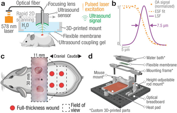
The large‐scale optoacoustic microscopy (LSOM) system and 3D‐printed mount for non‐invasive, label‐free, longitudinal monitoring of wound healing in the dorsal skin. a) Short‐pulsed laser light (578 nm) is focused through an ultrasound sensor into the skin, where it generates ultrasound waves following local optical absorption. The sensor is rapidly scanned over the dorsal skin to generate volumetric, high‐resolution images. b) Resolution characterization using an aluminum edge target showcases capillary‐level resolution of 7.5 µm measured as full‐width‐at‐half‐maximum (FWHM) of the line spread function (LSF). Orange dots – measured OA signal, dashed orange line – edge spread function (ESF) as logistic fit to OA data, solid purple line – LSF obtained by numerical differentiation of ESF. See also Experimental Section. c) Full‐thickness 5 mm excisional wounds in the dorsal skin of SKH1 mice were created. Inset showing freshly excised wounds (* indicates location of black marker used to match wound locations to pre‐wound imaging area). d) A 3D‐printed, custom‐made dorsal imaging mount stabilizes the mouse skin during imaging while providing a flat 11 × 30 mm² imaging area (see also Figure S3 and Video S1, Supporting Information).