Figure 3.

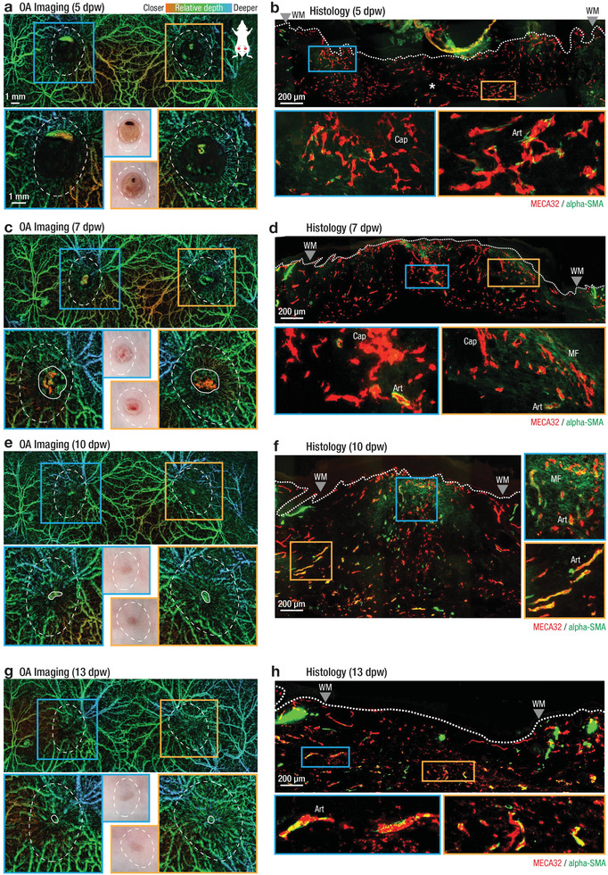
Observation of large‐scale angiogenesis and remodeling during cutaneous wound healing over a two‐week period using non‐invasive, label free LSOM and the corresponding histology in SKH1 mice. a,c,e,g) In vivo LSOM images of wounded dorsal skin at 5,7,10, and 13 dpw. Top panels provide a large‐scale overview of the dorsal vascular network, while bottom panels show a high‐resolution scan concentrating on the left (blue box and outline) and right (orange box and outline) wound regions. Middle inserts show gross appearance of wounds as digitally photographed. White dashed lines indicate approximate location of original wound area (see Figure S9, Supporting Information). White solid lines indicate border of wound edge vasculature. Images are color‐coded for relative vessel depth. b,d,f,h) Side view of excisional wounds at 5, 7, 10, and 13 dpw. Tissues were sectioned and stained using antibodies against MECA‐32 for endothelial cells (red) and α‐SMA for vascular smooth muscle cells and myofibroblasts (green); mature vessels show MECA‐32/α‐SMA co‐localization (yellow). Myofibroblasts are α‐SMA positive and MECA‐32 negative (faint green in (d,f). At 5 dpw, angiogenesis is limited to small caliber vessels near the original wound margins (a, white dashed lines), where histologically highly branched, sprouting, superficial capillaries (b, blue box), deep arterioles near the wound edge (b, orange box) and little vasculature in the wound center (b, white asterisk) were observed. Wound vasculature begins aligning toward the wound center at 7 dpw (c), also visible histologically as maturing, aligned arterioles at the wound edge surrounded by vascular smooth muscle cells (d, orange box). Macroscopic wound closure is complete at 10 dpw (e, photo inserts) with vascular remodeling ongoing, visible as more aligned mature vessels in both in vivo LSOM images (e) and histologically (f, orange box). At 13 dpw, angiogenesis is complete, with the entire wound closed (g), and histologically with mature vessels visible at the wound edge (h, blue box) and in the wound center (h, orange box). See Figure S10, Supporting Information, for additional wound staining and Figure S11, Supporting Information, for separate MECA‐32 and α‐SMA channel images. Art – arterioles; Cap – capillaries; WM – wound margins, MF – myofibroblasts.
