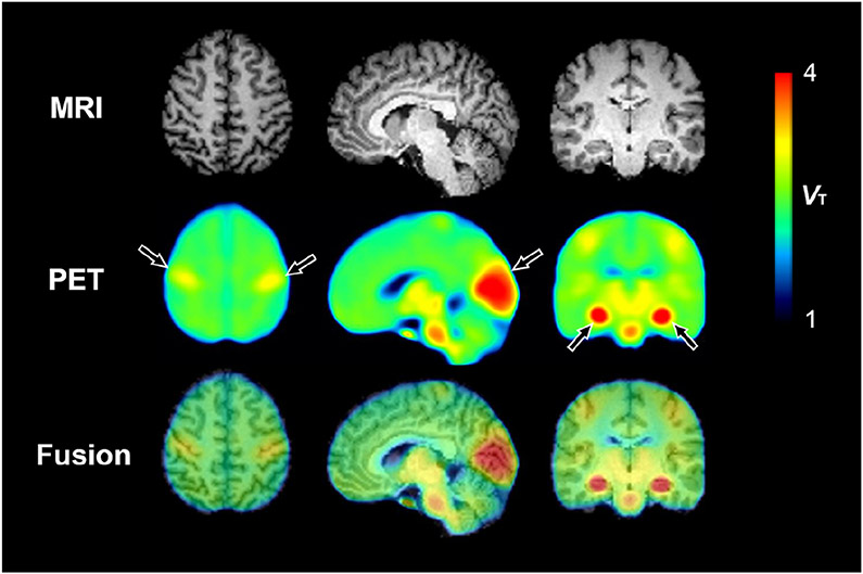
Fig. 2.
Parametric total distribution volume (VT) images calculated by the Logan graphical analysis method. MRI is from a representative participant, and PET images are averaged from 20 scans in 10 participants. Arrows represent notable [11C]PS13 binding in the pericentral cortex, occipital cortex, and hippocampus

