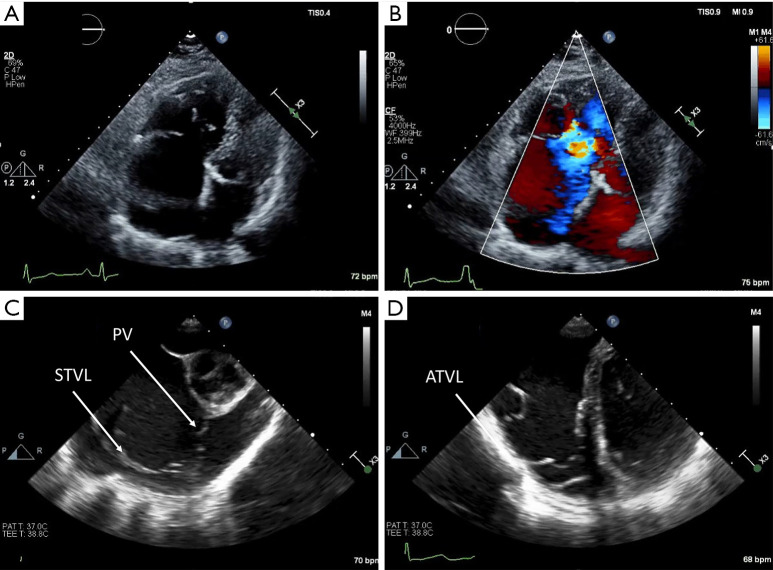
Figure 7.
Ebstein anomaly. Demonstrates a case of Ebstein anomaly on transthoracic echocardiography. (A) Apical four-chamber view showing right ventricular dilation and apical displacement of the tricuspid annulus due to restriction of the septal leaflet and a larger, redundant anterior leaflet. (B) Tricuspid regurgitation due to the dysmorphic valve is visualized on color Doppler. (C) Demonstrates the severity of valve abnormality in this case with the septal tricuspid valve leaflet closely juxtaposed to the pulmonary valve. (D) Demonstrates the classic ‘Sail’ morphology of the redundant anterior tricuspid leaflet in Ebstein anomaly. ATVL, anterior tricuspid valve leaflet; PV, pulmonary valve; STVL, septal tricuspid valve leaflet.

