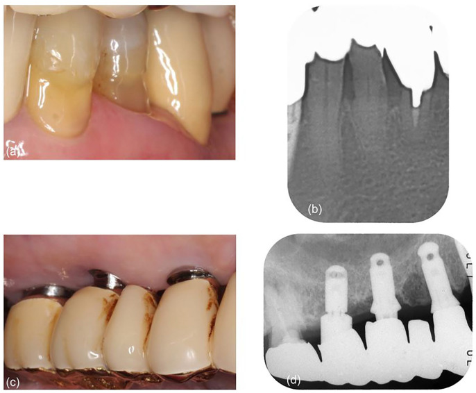
Figure 2.
Five-year follow-up: (a) gingiva is receded and tightened around the mandibular left first premolar; (b) section of panoramic radiograph shows the increase of bone density around the first premolar; (c) peri-implant mucosal recession is observed around the maxillary right molar implants. The margin of the superstructure is exposed to be supragingival; and (d) bone density is increased around the implants.

