
Figure 5. Histopathology of kidney in 12-month-old Arsk-deficient mice.
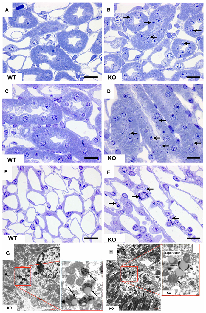
Toluidine blue-stainings of semi-thin sections of Arsk-deficient mice kidneys exhibit dense bodies (arrows) in the thick ascending limbs of the inner stripe of the outer medulla (B,D) as well as in intermediate tubules of the inner medulla (F); the respective WT controls are shown on the left (panels A, C and E). In electron microscopy (G,H), the equivalent of these dense bodies (arrows) appeared as lipofuscin-related material exclusively in the kidney of Arsk-deficient mice. Scale bars: 20 μm (light microscopy); 5 μm (electron microscopy).