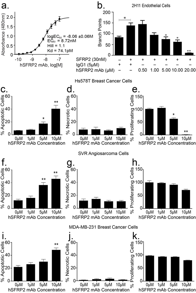
Figure 3: Humanized SFRP2 mAb in vitro activity.
(a) Humanized SFRP2 mAb binds recombinant human SFPR2 protein (SFRP2) with high affinity. Concentration-response curve showing the 480 nm absorbance measured after binding increasing concentrations of hSFRP2 mAb to a preset concentration of 1 μM SFRP2 in an ELISA assay (n=16). EC50: half-maximal effective concentration; Kd: equilibrium dissociation constant; Hill: Hill coefficient. (b) Bar graph showing the effects of SFRP2 and hSFRP2 mAb on 2H11 endothelial tube formation. 2H11 cells were incubated and either treated with IgG1 control only (5 μM), or IgG1 (5μM) + SFRP2 protein (30nM), or a combination of SFRP2 (30 nM) and hSFRP2 mAb (from 0.5 to 10μM). n=4 *: p≤ 0.05; **: p≤ 0.001. (c-h) Bar graphs showing the effects of increasing concentrations of hSFRP2 mAb (0 to 10 μM) on apoptosis (c, f, i), necrosis (d, g, j), and proliferation (e, h, k), in Hs578T breast cancer cells (c-e), and SVR angiosarcoma cells (f-h), and MDA-MB-231 cells (i-k). *: p ≤ 0.05; **: p ≤ 0.001. Proliferation was measured using Cyquant®, while apoptosis and necrosis were measured using Annexin V and propidium iodide. Results are a compilation of 3 independent experiments containing 4 wells each (n=12) for Hs578T and SVR; and 2 experiments containing 4 repeats for MDA-MD-231 (n=8).

