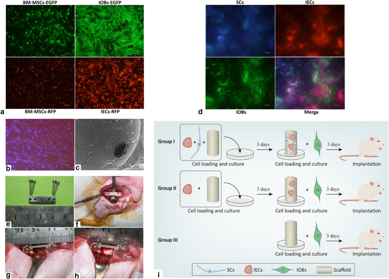
Fig. 4.
Prevascularized scaffolds loaded with labeled cells were applied in rat models. a BM-MSCs, IOBs expressing EGFP, and IECs expressing RFP (scale bar = 100). b SCs labeled with Hoechst 33342 (scale bar = 100). c SCs, IECs, and IOBs adhered and grew on β-TCP scaffolds observed by SEM at day 6 post-seeding. d Labeled SCs, IOBs-EGFP, and IECs-RFP grew on the β-TCP scaffolds to make prevascularized TEBGs (scale bar = 100) at day 6 post-seeding. e The internal fixation with steel plate applied in rat operations. f Femur exposed and drilled for internal fixation. g Six-millimeter-long defect of the middle femur and the steel plate implanted for internal fixation. h Prevascularized TEBG implanted into the femur defect. i The design of the time schedule and group setting during the experiment

