Fig. 4.

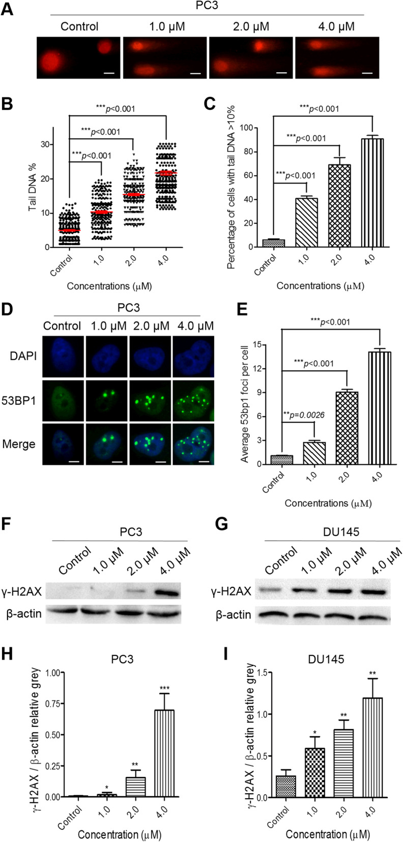
PL treatment triggers strong DNA damage and provokes an intense DDR in CRPC PC3 cells. A The data from comet assays show that PL treatment triggered strong DNA damage in a concentration-dependent manner. Scale bar, 100 μm. B and C, The percentages of tail DNA in PL-treated PC3 cells (B) and the number of PL-treated PC3 cells containing more than 10% tail DNA (C) were measured. Cells were treated with the indicated concentration of PL (1.0, 2.0, or 4.0 μM) for 48 h, and ≥ 500 cells were examined in each group. D The IF data show that PL treatment provoked an intense DDR in a concentration-dependent manner. Scale bar, 10 μm. E Quantification of (D). Cells were treated with the indicated concentration of PL (1.0, 2.0, or 4.0 μM) for 48 h, and ≥ 200 cells were examined in each group. F and G Western blotting of γ-H2AX in control (0.01% DMSO) and PL-treated PC3 (F) and DU145 (G) cells. Cells were treated with the indicated concentration of PL (1.0, 2.0, or 4.0 μM) for 48 h and were subjected to Western blotting using an antibody against γ-H2AX. β-actin was used as a loading control. H and I, Quantification of (F) and (G), respectively. Values represent the mean ± SD of at least three independent experiments. The statistical significance was calculated using the unpaired student’s two-tailed t-test with the p-values (*p < 0.05, **p < 0.01, ***p < 0.001)
