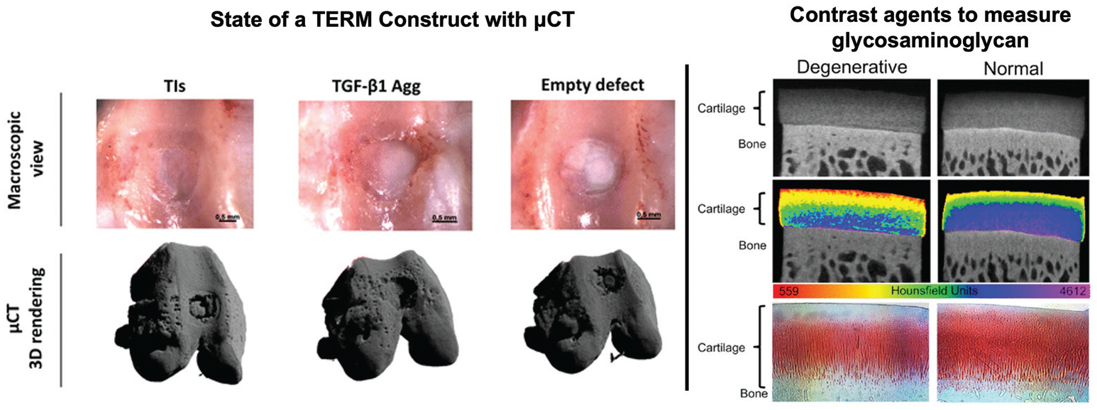
Fig. 3.

Computed tomography: Macroscopic view (top row) of TERM treated (left, middle) and untreated (right) osteochondral defects. μCT was used to render the osteochondral defect and subchondral bone to assess state of the TERM constructs and de novo tissue production. Figure reproduced with permission from Springer Nature (2018).39 (Right) A cationic contrast agent that is sensitive to glycosaminoglycan distribution in degenerated and normal cartilage. The contrast agent is attracted to the strong negative charge of glycosaminoglycans and increases radiopacity regions with high glycosaminoglycan concentration. This demonstrates how contrast agents can be used to assess the presence of biomaterials. Figure reproduced with permission from Elsevier (2018).53
