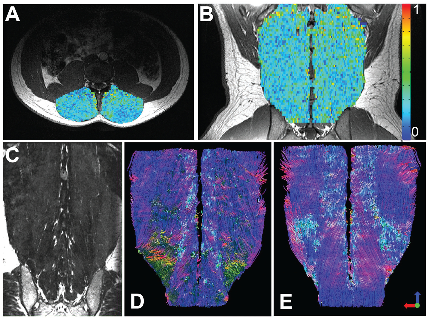
Fig. 5.

Diffusion tensor MRI: Axial (A) and coronal (B) fractional anisotropy maps overlaid on structural images of the lumbar paraspinal muscles, demonstrating the variance in tissue microstructural properties throughout a normal muscle. These maps can be used to assess microstructural organization of a TERM construct. In a coronal structural MRI scan (C) it is difficult to assess the 3D orientation of the paraspinal muscle fibers or assess fiber length. However, using tractography the orientation and length of the paraspinal muscles fibers can be measured. This technique can be used to assess how well a TERM construct is aligned and integrating with local tissue. Figure reproduced with permission from John Wiley and Sons (2020).115
