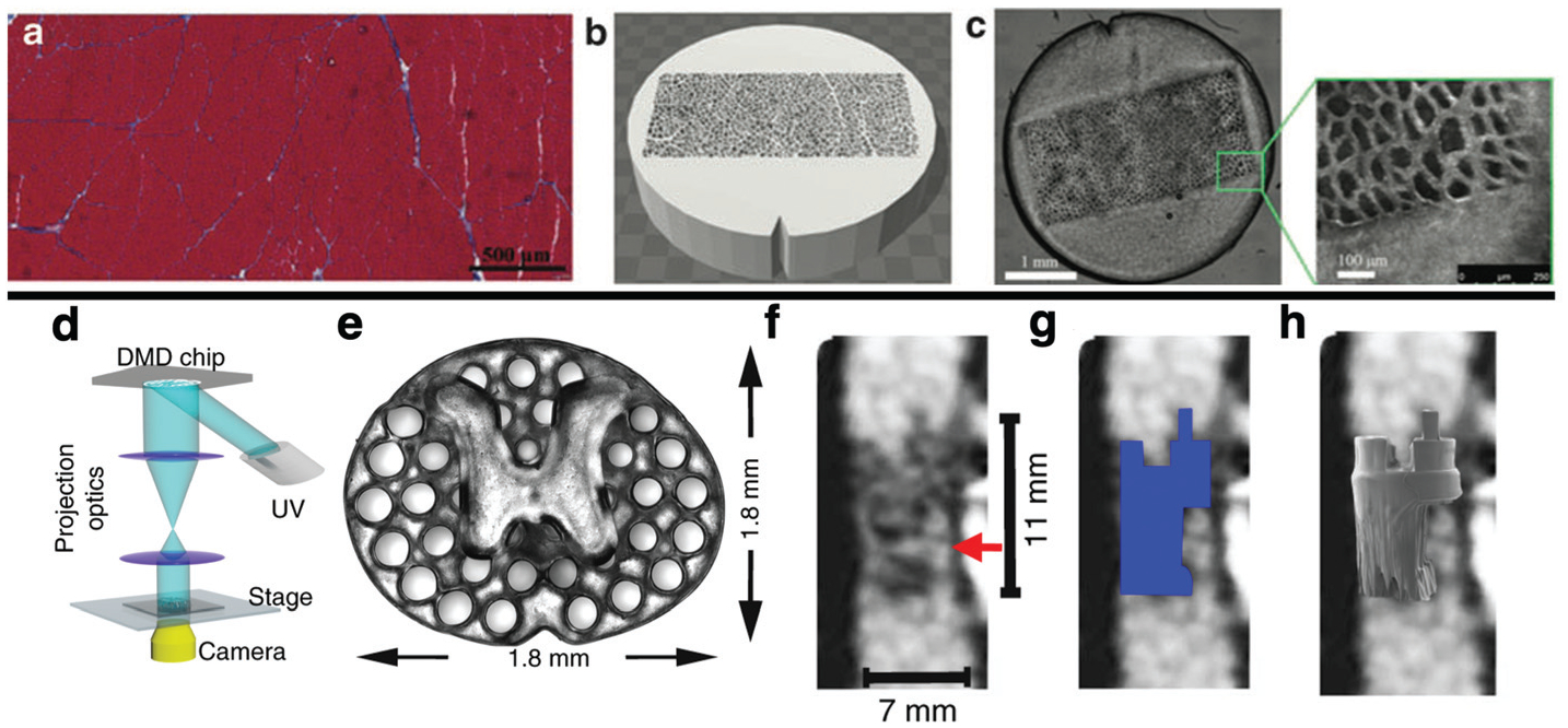
Fig. 6.

3D printing can be used to validate imaging techniques (Top). Histology of normal muscle (a) was used to inform the design of a phantom (b) with known geometric properties, which could be 3D printed (c) and scanned using MRI. This approach was used to relate measurements made using diffusion tensor MRI to known microstructural properties of the phantom.137 Biomedical imaging can also be used to inform the design of TERM constructs. A light based 3D printer (d) was used to print a scaffold (e) with x–y geometry informed by the axial distribution of white matter and grey matter in the spinal chord. Structural MRI of a complete spinal cord injury (f) can be used to inform the 3D geometry of the TERM scaffold (g), which can be precisely printed (h) to the precise dimensions of a patient’s lesion.139
