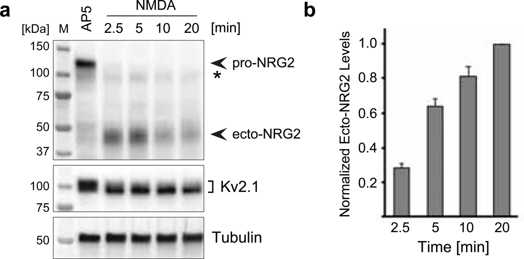
Fig. 1. Rapid NRG2 ectodomain shedding after NMDAR stimulation in cultured hippocampal neurons.
(a) Representative Western blot of whole-cell lysates prepared from AAV-transduced hippocampal neurons expressing wt NRG2 and treated with 100 μM AP5 or with 50 μM NMDA for the indicated times. The blot was probed with anti-NRG2 (Ab 7215) raised against its extracellular domain (top). It illustrates the rapid decrease of pro-NRG2 signals at ~120 kDa apparent molecular mass in response to NMDAR activation, and a concomitant increase in ecto-NRG2 levels at ~45 kDa apparent molecular mass. The weak band marked with an asterisk (*) is only detected in transduced neurons and likely reflects immature NRG2 protein. The blot was re-probed with anti-Kv2.1 to show the characteristic downward electrophoretic mobility shift of Kv2.1 protein in response to NMDAR stimulation (middle). Tubulin signals served as reference (bottom). (b) Corresponding ELISA measurements of ecto-NRG2 accumulation in conditioned cell culture supernatants demonstrating the parallel increase in levels of soluble ecto-NRG2. Data are normalized to NMDA at 20 min (by which time pro-NRG2 protein signals were consistently undetectable in Western blots) and represent the mean ± SEM from 3 independent experiments.

