. 2021 May 21;41(4):E1064–E1081. doi: 10.1148/rg.2021200181

Figure 12.

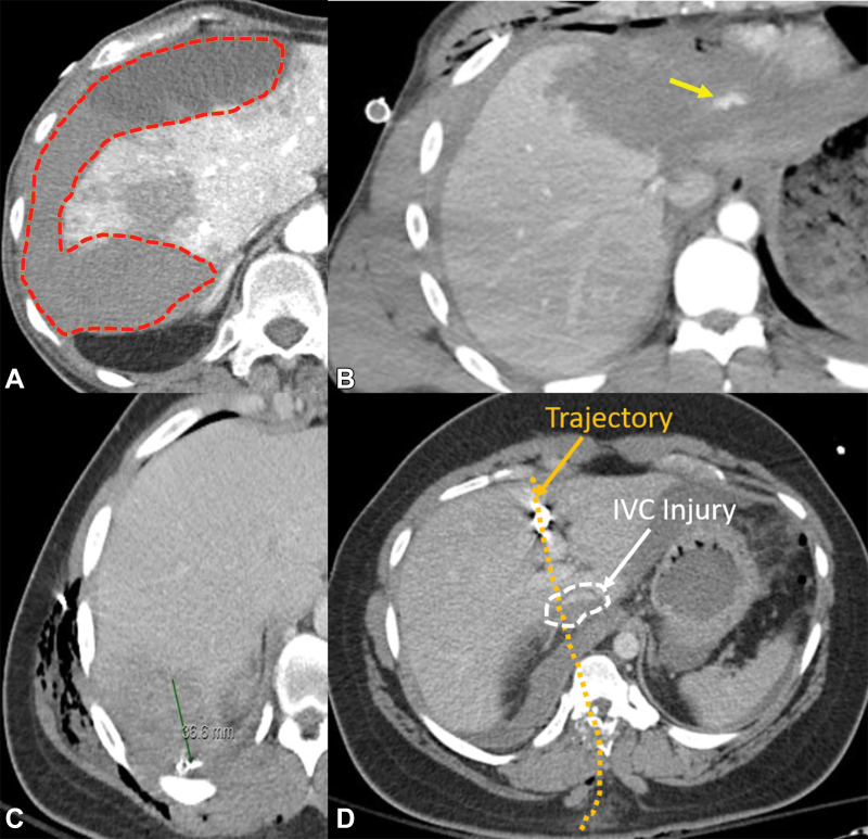
Description of various imaging features that may help in grading liver injury. Axial CT images depict hematoma (orange outline in A), active extravasation (arrow in B), laceration (green line indicating ruler in C), and juxtavenous injury (white arrow and dashed circle in D). The trajectory is superimposed (orange arrow and orange dotted line in D). IVC = inferior vena cava.

Description of various imaging features that may help in grading liver injury. Axial CT images depict hematoma (orange outline in A), active extravasation (arrow in B), laceration (green line indicating ruler in C), and juxtavenous injury (white arrow and dashed circle in D). The trajectory is superimposed (orange arrow and orange dotted line in D). IVC = inferior vena cava.