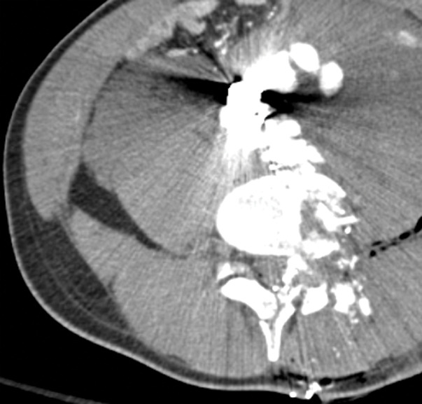
Figure 16c.
Direct and indirect signs of vascular injury in four patients. (a) Axial contrast-enhanced CT image of the abdomen and pelvis in a 34-year-old woman with a gunshot injury to the right flank demonstrates hematoma (circle) abutting the right renal artery (arrow) with vasospasm, which is suggestive of low-grade injury. (b) Axial contrast-enhanced CT image of the abdomen and pelvis in a 25-year-old man with a stab wound to the posterior flank from a long knife demonstrates right renal laceration with perinephric hematoma and a focal outpouching from the right renal artery (arrow), which are consistent with pseudoaneurysm. (c, d) Axial contrast-enhanced CT images of the abdomen and pelvis in a 19-year-old patient with a gunshot injury to the left back demonstrate hematoma in the retroperitoneum with vascular injuries to the aorta and inferior vena cava (obscured by streak from bullets) with active extravasation (arrow in d), suggesting vascular injury. (e) Axial CT image in a 27-year-old man with a gunshot injury to the right lateral abdomen (entry wound shown with solid orange oval) depicts a trajectory superimposed on the image (arrow) that traverses through the aorta and inferior vena cava, causing an arteriovenous fistula (dashed yellow oval).

