. 2021 May 21;41(4):E1064–E1081. doi: 10.1148/rg.2021200181

Figure 18.

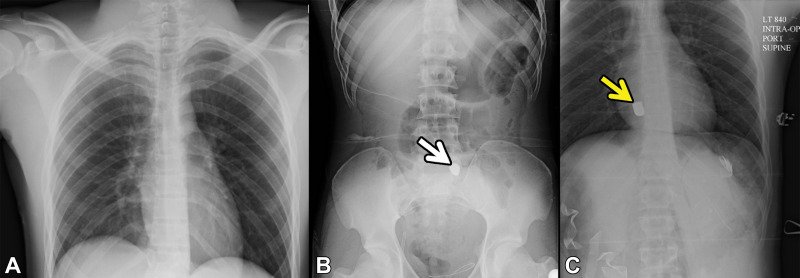
Bullet embolism in a 19-year-old patient who presented to the ED with gunshot trauma and hemodynamic instability. A, B, Anteroposterior radiographic survey images obtained in the trauma bay before exploratory laparotomy demonstrate a bullet projecting over the sacrum (arrow in B ). C, Supine radiograph that was acquired intraoperatively to reveal the position of the bullet after it could not be found in the pelvis shows that the bullet is embolized to the right ventricle (arrow). This imaging finding is direct evidence of vascular injury.

Bullet embolism in a 19-year-old patient who presented to the ED with gunshot trauma and hemodynamic instability. A, B, Anteroposterior radiographic survey images obtained in the trauma bay before exploratory laparotomy demonstrate a bullet projecting over the sacrum (arrow in B ). C, Supine radiograph that was acquired intraoperatively to reveal the position of the bullet after it could not be found in the pelvis shows that the bullet is embolized to the right ventricle (arrow). This imaging finding is direct evidence of vascular injury.