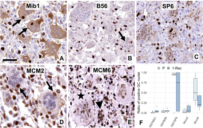
FIGURE 2.
Expression of “general” proliferation markers i.e. Ki67 (A–C) and MCM-complex proteins (D–E) in multinucleated GCs. Mib1 (A) and B56 (B) antibodies showed occasional nuclear immunoreactions (arrows), while the SP6 clone resulted in usually weaker, but a widespread Ki67 positivity in GCs. Cytoplasmic Mib1 positivity in GCs was validated by negativity in several mononuclear cells. MCM2 reaction (arrows) was also rare in GCs (D), while that of MCM6 was rather frequent (E) and obviously more pronounced in smaller (arrowhead), than larger GCs (asterisk). Scale bar represents 40 μm on A, 50 μm on (B,C,E), and 30 μm on (D). Boxplot of the ratio of immunopositive GC nuclei vs. all GC nuclei (F) in primary (P) and first recurrent (1-Rec) GCTB cases.

