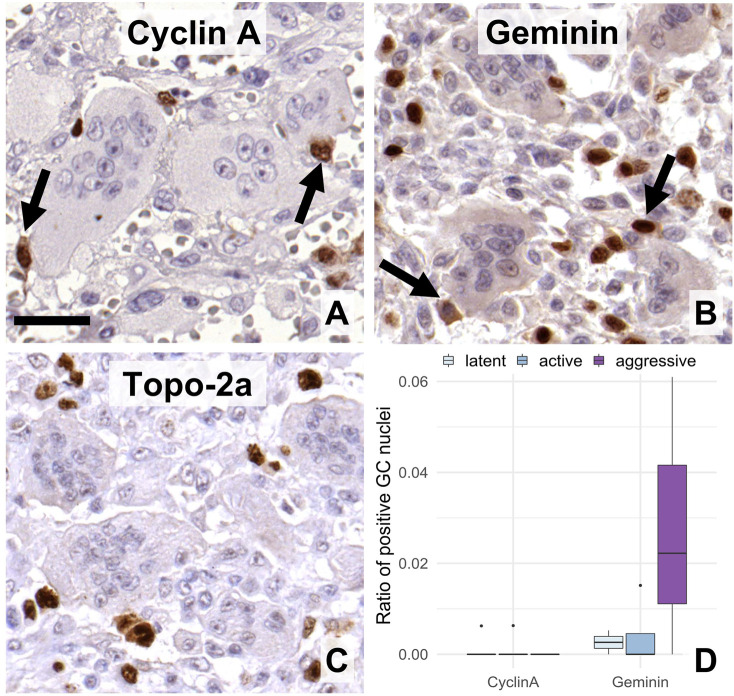
FIGURE 4.
Expression of post-G1 (S-G2-M)-phase cell cycle markers in GC. Cyclin A (A), geminin (B), and topoisomerase-2a (C) were practically detected only in the mononuclear cells. Arrows show immunopositive mononuclear cells of close association with multinucleated GCs. Scale bar: 30 μm for all images. Very rare geminin positive cells were somewhat more frequent in agressive grade tumors vs. the other groups ((D), boxlot).

