
Figure 2:
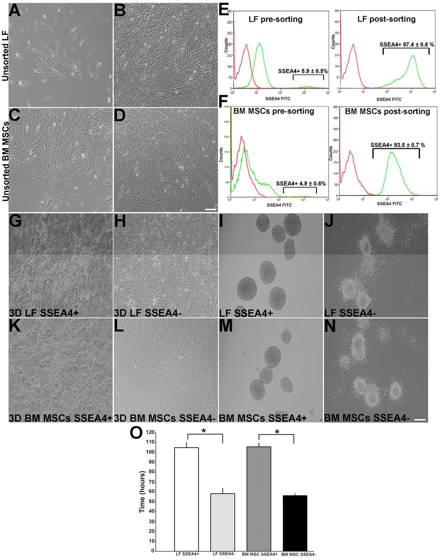
Culture of LF and BM MSC cells. (A): Cultured human LFs at day 6. (B): LFs near confluence at day 15. (C): BM MSCs at day 2. (D): BM MSCs near confluence at day 7. (E): LFs’ expression of SSEA4 pre-sorting and SSEA4 post-sorting. (F): BM MSCs pre-sorted and post-sorted for SSEA4 (green line indicates antibody expression and red line isotype control). Sorted LFs. (G): LF SSEA4+ cells in 3D cultures expressing cells with round compact morphology. (H): LF SSEA4− cells expressing fibroblast morphology. (I): LF SSEA4+ spheres. (J): LF SSEA4− spheres with outgrowth at day 7. (K): BM MSC SSEA4+ cells grown in 3D culture expressing round compact morphology at day 5. (L): BM MSC SSEA4− cells expressing fibroblast morphology day 5. (M): BM MSC SSEA4+ spheres at day 6. (N): SSEA4− spheres with outgrowth. (O): Doubling time of SSEA4+ LF and BM MSC cells are higher than those in the SSEA4− groups. Scale bar 20 μm. The data represent the mean ± SEM from 6 experiments. Statistical analysis was performed using Two tailed Student’s t test (*p < 0.05). Abbreviations: LFs, limbal fibroblasts; BM MSCs, bone marrow mesenchymal stem cells; SSEA4, stage-specific embryonic antigen 4.