
Figure 4:
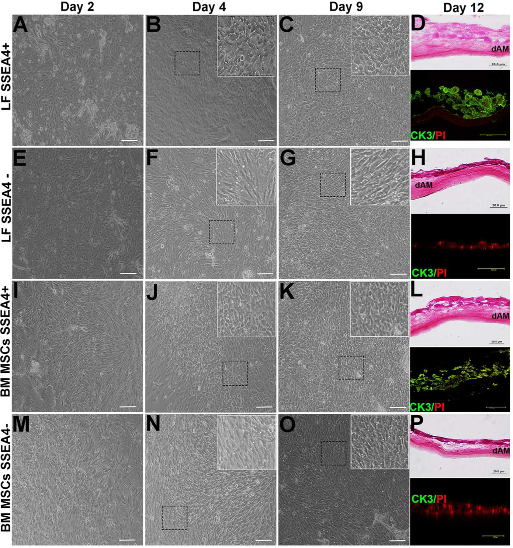
Differentiation of LFs, BM MSCs. (A): LF SSEA4+ cells at day 2. (B): LF SSEA4+ cells at day 4, cells appear to be round and compact (inset) (C): LF SSEA4+ cells appear epithelia-like at day 9 (inset). (D): Cross section of SSEA4+ LFs grown on dAM and stained with H&E at day 12 and CK3 (green) positive cells. (E): LF SSEA4− cells at day 2. (F): At day 4, LF SSEA4− cells appear fibroblast-like (inset). (G): At day 9, LF SSEA4− cells appear fibroblast-like (inset). (H): Cross section of SSEA4− LFs grown on dAM and stained with H&E at day 12 and CK3 (green) negative cells. (I): BM MSCs SSEA4+ 2 days after plating. (J): At day 4, cells appear to be round and compact in form (inset). (K): BM MSCs SSEA4+ appear round epithelial-like cell morphology at day 9 (inset). (L): Cross section of SSEA4+ BM MSCs grown on dAM and stained with H&E at day 12 and CK3 (green) positive cells. (M): BM MSC SSEA4− cells at day 2. (N): At day 4, cells appear fibroblast-like (inset). (O): BM MSC SSEA4− cells appear fibroblast-like at day 9 (inset). (P): Cross section of SSEA4+ LFs grown on dAM and stained with H&E at day 12 and CK3 (green) negative cells. Scale bar 20 μm. Inset images 40 × magnifications. Abbreviations: LFs, limbal fibroblasts; BM MSCs, bone marrow mesenchymal stem cells; SSEA4, stage-specific embryonic antigen 4; dAM, denuded amniotic membrane; H&E, hematoxylin and eosin; CK3, cytokeratin 3; PI, Propidium iodide (nuclear stain).