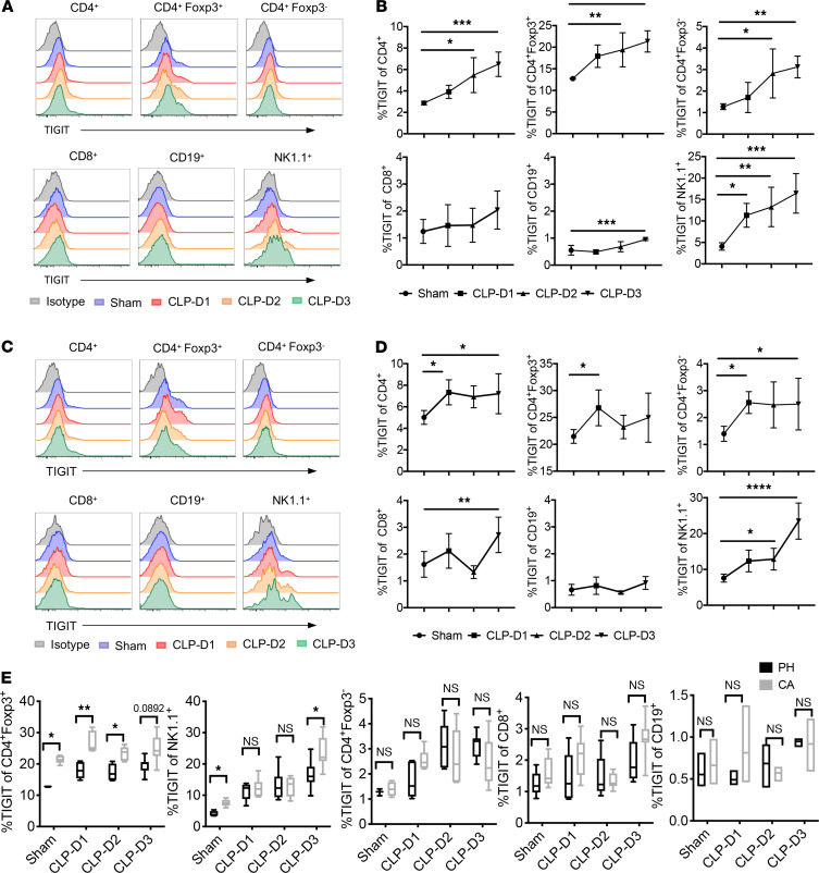
Figure 1. TIGIT is upregulated on splenic lymphocytes isolated from PH septic mice and septic mice with preexisting malignancy (CA).
B6 mice were injected with LLC and monitored for 3 weeks. PH and CA mice were subjected to CLP (n = 22/group) or sham surgery (n = 5/group). Mice were sacrificed on days 1, 2, and 3 after CLP and TIGIT expression on splenic immune cells was measured. (A and C) Representative flow histograms showing TIGIT expression on the indicated lymphocyte populations isolated from either PH or CA septic mice. Plots were gated on CD4+, CD4+Foxp3+/–, CD8+, NK1.1+, and CD19+ cells, respectively. (B and D) Summary data of the percentage of TIGIT+ lymphocytes isolated from either PH or CA septic mice. (E) TIGIT expression on each cell subset was compared between PH and CA mice at different time points. Groups were compared with 2-way ANOVA with Tukey’s post hoc test. *P ≤ 0.05, **P ≤ 0.01, ***P ≤ 0.001. PH, previously healthy; CA, cancer; LLC, Lewis lung carcinoma; CLP, cecal ligation and puncture; TIGIT, T cell Ig and ITIM domain.

