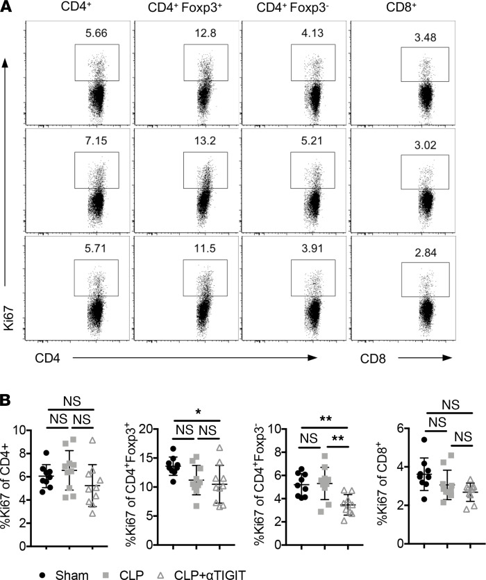
Figure 7. Decreased lymphocyte loss in anti-TIGIT–treated CA septic mice was not associated with increased proliferation.
CA mice were subjected to CLP and either treated with anti-TIGIT mAb or the same volume of PBS as described above. Mice were subjected to sham surgery as a control. Spleens were collected for intracellular Ki67 staining. Data were derived from 2 independent experiments. n = 9–10/group. (A) Representative flow cytometry plots of Ki67 expression on total CD4+ T cells, Treg, Tconv, and total CD8+ T cells in the 3 groups. (B) Data summary of Ki67 percentages on different cell subsets. Groups were compared with 2-way ANOVA with Tukey’s post hoc test. *P ≤ 0.05, **P ≤ 0.01. CA, cancer; CLP, cecal ligation and puncture; TIGIT, T cell Ig and ITIM domain; Tconv, T conventional cell.

