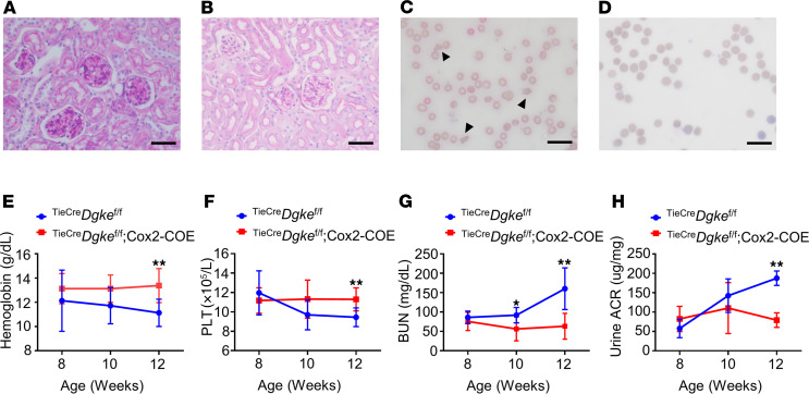
Figure 4. Overexpression of Cox2 in endothelial cells rescues the phenotype of endothelial specific Dgke-knockout mice.
(A) Representative bright-field microscopy images of glomeruli of PAS-stained Tie2CreDgkefl/fl mouse kidneys and (B) Tie2CreDgkefl/fl Cox2-COE mouse kidneys at 12 weeks of age. The occlusion of the glomerular capillaries is rescued in Tie2CreDgkefl/fl Cox2-COE mice. (C) Smears of blood from Tie2CreDgkefl/fl knockout mice and (D) Tie2CreDgkefl/fl Cox2-COE mice at 3 months of age. Schistocytes are visible in Tie2CreDgkefl/fl knockout mice (arrowheads) and are absent in Tie2CreDgkefl/fl Cox2-COE mice. Scale bars are 50 μm. (E–H) Serum hemoglobin, circulating PLT, BUN, and urine ACR in Tie2CreDgkefl/fl and Tie2CreDgkefl/fl Cox2-COE mice at 8, 10, and 12 weeks of age. Data are presented as mean ± SD. *: P < 0.05, **: P < 0.01 by Student’s t test. n = 6 mice per group.

