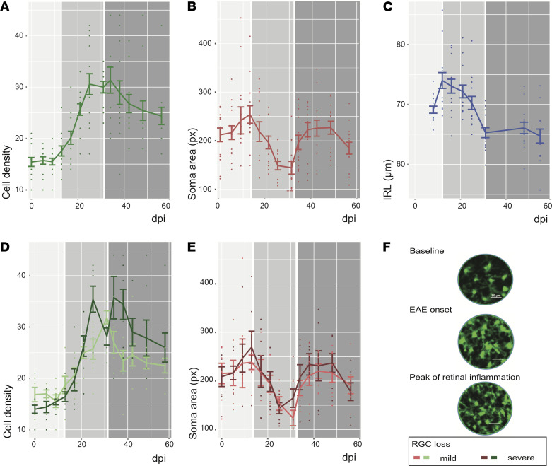
Figure 1. Longitudinal in vivo imaging in EAE.
(A and B) Preonset (light gray 0–13 dpi), the cell density (A) and soma area (B) of CX3CR1 GFP+ cells increase. Whereas the cell density of CX3CR1 GFP+ cells further increases during the acute phase (13–30 dpi; medium gray) and decreases in the chronic phase (31–56 dpi; dark gray), the soma area of CX3CR1 GFP+ cells decreases in the acute phase and then peaks again the chronic phase. (C) The IRL thickness increases before onset, drops dramatically during the acute phase, and stabilizes in the chronic phase of EAE. (D and E) Eyes with severe RGC loss compared with eyes with mild RGC loss at 56 dpi showed a higher cell density of CX3CR1 GFP+ cells 25 dpi and 38 dpi (D) and a less relevant drop of CX3CR1 GFP+ cell soma area (E). (F) Cell density and soma area changes of CX3CR1 GFP+ cells are qualitatively visible by CSLO (please note: images falsely colorized). For A–E, data are shown as mean ± SEM; experiment included eyes of 7 mice.

