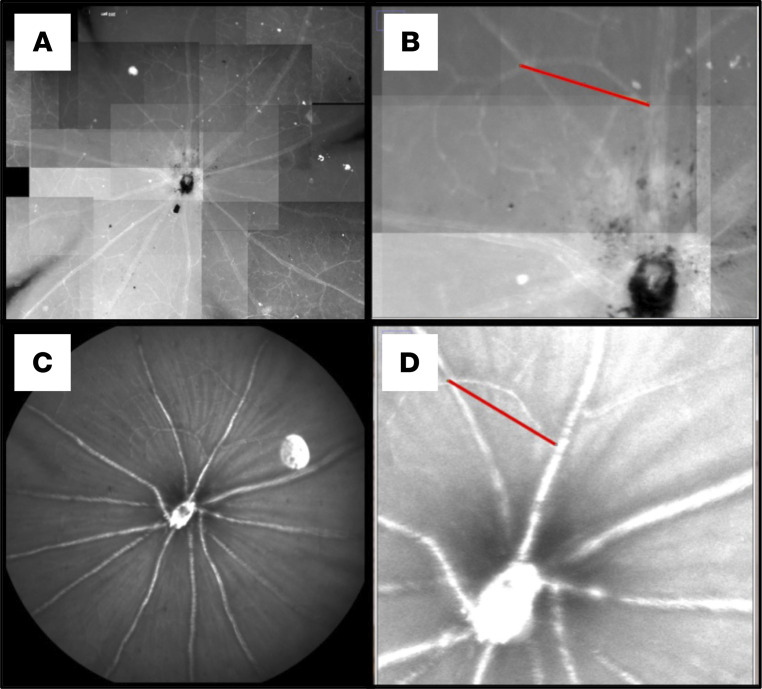
Figure 5. Reliability of CSLO measurements.
(A and B) Microscopy image from a retinal flat mount. (C and D) The same retina, imaged in vivo through CSLO centered on the optic nerve head, one day prior to eye extraction. The red lines on B and D show measurements of the same arterial branch in vitro (B) and in vivo (D).

