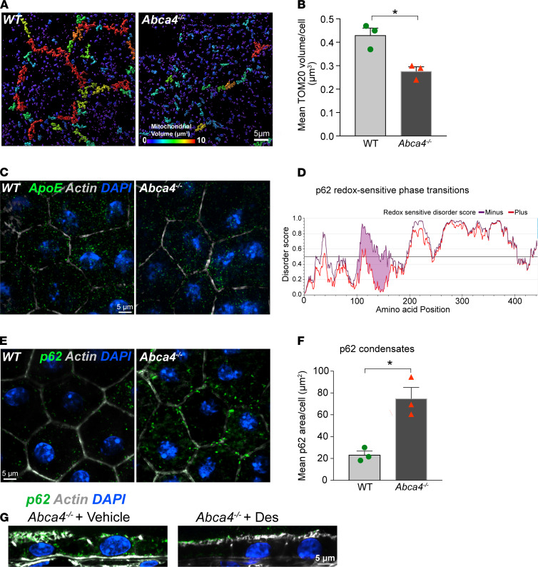
Figure 6. Mitochondrial injury and redox-sensitive protein phase separation in Abca4–/– mouse RPE.
(A) Volume reconstructions of TOM20-stained mitochondria in WT and Abca4–/– mouse RPE flatmounts. Warmer colors in the color bar indicate larger volumes. (B) Quantification of mean TOM20 volumes from images in A. Mean ± SEM, *P < 0.05, n = 3 mice per genotype. (C) ApoE immunostaining (green) in WT and Abca4–/– mouse RPE flatmounts. (D) IUPRED2 redox-driven disorder predictions for mouse p62. (E) p62 condensates (green) in WT and Abca4–/– RPE flatmounts. (F) Areas of p62 aggregates. Mean ± SEM; *P < 0.05; n = 3 mice per genotype. (G) p62 (green) in RPE in Abca4–/– mice treated with vehicle or desipramine. (C, E, and G) Nuclei, DAPI (blue); actin, phalloidin (gray).

