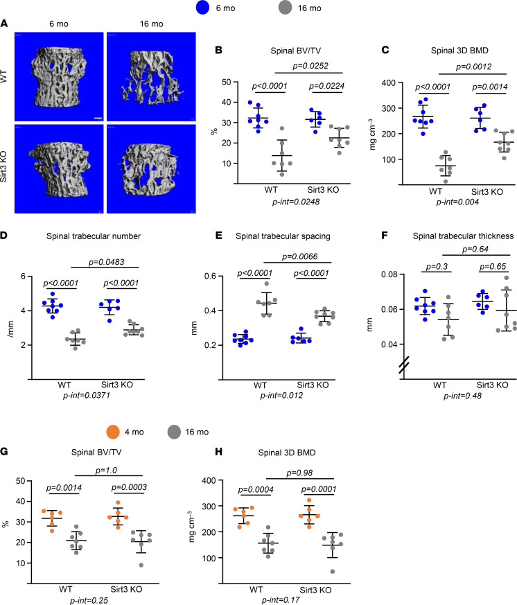
Figure 2. Deletion of Sirt3 attenuates age-associated trabecular bone loss.
(A–F) Imaging and quantification of vertebral bones from female Sirt3-KO mice and WT littermates by micro-CT after sacrifice (n = 7–10 animals/group). Representative images of trabecular bone (A) and bone volume per tissue volume (BV/TV), bone mineral density (BMD), and microarchitecture of trabecular bone in L5 (B–F). Scale bar: 100 μm. (G and H) BV/TV and BMD from male Sirt3-KO mice and WT littermates in L5 bones by micro-CT (n = 5–7 animals/group). Data are presented as ± SD. P values were determined using 2-way ANOVA. Interaction terms generated by 2-way ANOVA are shown below each graph.

